Nikon Small World Photomicrography Competition 2012
Advertisements:
Most people know Nikon as a purveyor of pro and consumer-grade digital cameras. But the company's expertise with optics bleeds over into related markets – it's one of the science community's major suppliers of microscopes. And each year the company asks the community to send it some of their favorite images of tiny objects. A panel of scientists and journalists have chosen the best of this past year's submissions, which Nikon has placed on its Small World site.
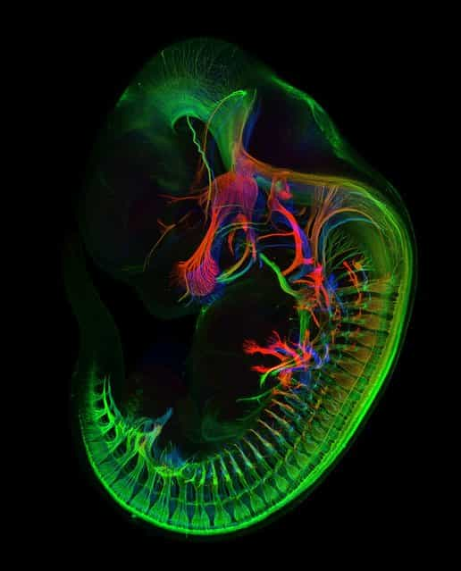
1st Place. [The blood-brain barrier in a live zebrafish embryo (20x)]. The blood-brain barrier plays a critical role in neurological function and disease. Drs. Peters and Taylor, developed a transgenic zebrafish to visualize the development of this structure in a live specimen. By doing so, this model proves that not only can we image the blood-brain barrier, but we can also genetically and chemically dissect the signaling pathways that modulate the blood-brain barrier function and development. To achieve this image, Peters and Taylor used a maximum intensity projection of a series of images acquired in the z plane. The images were first pseudo-colored with a rainbow palette based on depth so that the coloring scheme would be both visually appealing and provide spatial information. In doing so, Peters and Taylor captured an image that Peters says [not only captures the beauty of nature, but is also topical and biomedically relevant]. (Photo by Dr. Jennifer L. Peters & Dr. Michael R. Taylor, St. Jude Children’s Research Hospital Memphis, Tennessee, USA)
2nd Place. [Live newborn lynx spiderlings (6x)]. (Photo by Walter Piorkowski, South Beloit, Illinois, USA)
3rd Place. [Human bone cancer (osteosarcoma) showing actin filaments (purple), mitochondria (yellow), and DNA (blue) (63x)]. (Photo by Dr. Dylan Burnette, National Institute of Health (NIH) Bethesda, Maryland, USA)
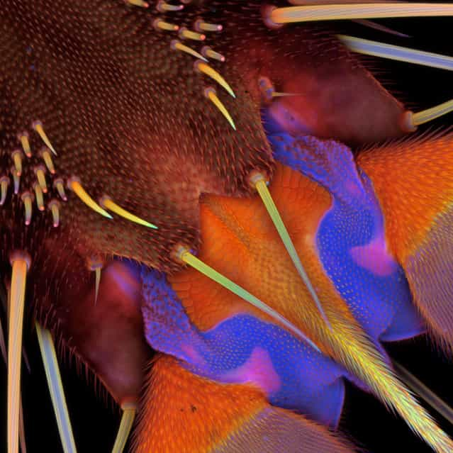
4th Place. [Drosophila melanogaster visual system halfway through pupal development, showing retina (gold), photoreceptor axons (blue), and brain (green) (1500x)]. (Photo by Dr. W. Ryan Williamson, Howard Hughes Medical Institute (HHMI), Ashburn, Virginia, USA)
5th Place. [Cacoxenite (mineral) from La Paloma Mine, Spain (18x)]. (Photo by Honorio Cócera-La Parra, University of Valencia, Museum of Geology, Department of Geology, Benetusser, Valencia, Spain)
6th Place. [Cosmarium sp. (desmid) near a Sphagnum sp. leaf (100x)]. (Photo by Marek Miś, Marek Mis Photography, Suwalki, Poland)
7th Place. [Eye organ of a Drosophila melanogaster (fruit fly) third-instar larvae (60x)]. (Photo by Dr. Michael John Bridge, University of Utah, HSC Core Research Facilities – Cell Imaging Lab, Salt Lake City, Utah, USA)
8th Place. [Pleurobrachia sp. (sea gooseberry) larva (500x)]. (Photo by Gerd A. Guenther, Düsseldorf, Germany)
9th Place. [Myrmica sp. (ant) carrying its larva (5x)]. (Photo by Geir Drange, Asker, Norway)
10th Place. [Brittle star (8x)]. (Photo by Dr. Alvaro Migotto, University of São Paulo, São Paulo, Brazil)
12th Place. [3D lymphangiogenesis assay. Cells sprout from dextran beads embedded in fibrin gel]. (Photo by Esra Guc, École Polytechnique Fédérale de Lausanne (EPFL), Lausanne, Vaud, Switzerland)
17th Place. [Stinging nettle trichome on leaf vein (100x)]. (Photo by Charles Krebs, Charles Krebs Photography, Issaquah, Washington, USA)
20th Place. [Embryos of the species Molossus rufus, the black mastiff bat. These images formed part of an embryonic staging system for this species]. (Photo by Dorit Hockman, University of Cambridge, Department of Physiology, Development and Neuroscience, Cambridge, United Kingdom)
19th Place. [Floral primordia of Allium Sativum (Garlic)]. (Photo by Dr. Somayeh Naghiloo, University of Tabriz, Department of Plant Biology, Faculty of Natural Sciences, Tabriz, East Azarbayedjan, Iran)
Honorable Mention. [Ptilota (red algae) (10x)]. (Photo by Dr. Arlene Wechezak, Anacortes, Washington, USA)
16th Place. [Fossilized Turitella agate containing Elimia tenera (freshwater snails) and ostracods (seed shrimp) (7x)]. (Photo by Douglas Moore, University of Wisconsin – Stevens Point, University Relations & Communications/Geology, Stevens Point, Wisconsin, USA)
Honorable Mention. [Snow crystal, illuminated with colored lights (5x)]. (Photo by Dr. Kenneth Libbrecht, California Institute of Technology (Caltech), Department of Physics, Pasadena, California, USA)
![Nikon Small World Photomicrography Competition 2012. 1st Place. [The blood-brain barrier in a live zebrafish embryo (20x)]. The blood-brain barrier plays a critical role in neurological function and disease. Drs. Peters and Taylor, developed a transgenic zebrafish to visualize the development of this structure in a live specimen. By doing so, this model proves that not only can we image the blood-brain barrier, but we can also genetically and chemically dissect the signaling pathways that modulate the blood-brain barrier function and development. To achieve this image, Peters and Taylor used a maximum intensity projection of a series of images acquired in the z plane. The images were first pseudo-colored with a rainbow palette based on depth so that the coloring scheme would be both visually appealing and provide spatial information. In doing so, Peters and Taylor captured an image that Peters says [not only captures the beauty of nature, but is also topical and biomedically relevant]. (Photo by Dr. Jennifer L. Peters & Dr. Michael R. Taylor, St. Jude Children’s Research Hospital Memphis, Tennessee, USA) Nikon Small World Photomicrography Competition 2012. 1st Place. [The blood-brain barrier in a live zebrafish embryo (20x)]. The blood-brain barrier plays a critical role in neurological function and disease. Drs. Peters and Taylor, developed a transgenic zebrafish to visualize the development of this structure in a live specimen. By doing so, this model proves that not only can we image the blood-brain barrier, but we can also genetically and chemically dissect the signaling pathways that modulate the blood-brain barrier function and development. To achieve this image, Peters and Taylor used a maximum intensity projection of a series of images acquired in the z plane. The images were first pseudo-colored with a rainbow palette based on depth so that the coloring scheme would be both visually appealing and provide spatial information. In doing so, Peters and Taylor captured an image that Peters says [not only captures the beauty of nature, but is also topical and biomedically relevant]. (Photo by Dr. Jennifer L. Peters & Dr. Michael R. Taylor, St. Jude Children’s Research Hospital Memphis, Tennessee, USA)](http://img.gagdaily.com/uploads/posts/app/2013/thumbs/00009fa7_medium.jpg)
![Nikon Small World Photomicrography Competition 2012. 2nd Place. [Live newborn lynx spiderlings (6x)]. (Photo by Walter Piorkowski, South Beloit, Illinois, USA) Nikon Small World Photomicrography Competition 2012. 2nd Place. [Live newborn lynx spiderlings (6x)]. (Photo by Walter Piorkowski, South Beloit, Illinois, USA)](http://img.gagdaily.com/uploads/posts/app/2013/thumbs/00009fa0_medium.jpg)
![Nikon Small World Photomicrography Competition 2012. 3rd Place. [Human bone cancer (osteosarcoma) showing actin filaments (purple), mitochondria (yellow), and DNA (blue) (63x)]. (Photo by Dr. Dylan Burnette, National Institute of Health (NIH) Bethesda, Maryland, USA) Nikon Small World Photomicrography Competition 2012. 3rd Place. [Human bone cancer (osteosarcoma) showing actin filaments (purple), mitochondria (yellow), and DNA (blue) (63x)]. (Photo by Dr. Dylan Burnette, National Institute of Health (NIH) Bethesda, Maryland, USA)](http://img.gagdaily.com/uploads/posts/app/2013/thumbs/00009fa2_medium.jpg)
![Nikon Small World Photomicrography Competition 2012. 4th Place. [Drosophila melanogaster visual system halfway through pupal development, showing retina (gold), photoreceptor axons (blue), and brain (green) (1500x)]. (Photo by Dr. W. Ryan Williamson, Howard Hughes Medical Institute (HHMI), Ashburn, Virginia, USA) Nikon Small World Photomicrography Competition 2012. 4th Place. [Drosophila melanogaster visual system halfway through pupal development, showing retina (gold), photoreceptor axons (blue), and brain (green) (1500x)]. (Photo by Dr. W. Ryan Williamson, Howard Hughes Medical Institute (HHMI), Ashburn, Virginia, USA)](http://img.gagdaily.com/uploads/posts/app/2013/thumbs/00009fa1_medium.jpg)
![Nikon Small World Photomicrography Competition 2012. 5th Place. [Cacoxenite (mineral) from La Paloma Mine, Spain (18x)]. (Photo by Honorio Cócera-La Parra, University of Valencia, Museum of Geology, Department of Geology, Benetusser, Valencia, Spain) Nikon Small World Photomicrography Competition 2012. 5th Place. [Cacoxenite (mineral) from La Paloma Mine, Spain (18x)]. (Photo by Honorio Cócera-La Parra, University of Valencia, Museum of Geology, Department of Geology, Benetusser, Valencia, Spain)](http://img.gagdaily.com/uploads/posts/app/2013/thumbs/00009fa3_medium.jpg)
![Nikon Small World Photomicrography Competition 2012. 6th Place. [Cosmarium sp. (desmid) near a Sphagnum sp. leaf (100x)]. (Photo by Marek Miś, Marek Mis Photography, Suwalki, Poland) Nikon Small World Photomicrography Competition 2012. 6th Place. [Cosmarium sp. (desmid) near a Sphagnum sp. leaf (100x)]. (Photo by Marek Miś, Marek Mis Photography, Suwalki, Poland)](http://img.gagdaily.com/uploads/posts/app/2013/thumbs/00009fa4_medium.jpg)
![Nikon Small World Photomicrography Competition 2012. 7th Place. [Eye organ of a Drosophila melanogaster (fruit fly) third-instar larvae (60x)]. (Photo by Dr. Michael John Bridge, University of Utah, HSC Core Research Facilities – Cell Imaging Lab, Salt Lake City, Utah, USA) Nikon Small World Photomicrography Competition 2012. 7th Place. [Eye organ of a Drosophila melanogaster (fruit fly) third-instar larvae (60x)]. (Photo by Dr. Michael John Bridge, University of Utah, HSC Core Research Facilities – Cell Imaging Lab, Salt Lake City, Utah, USA)](http://img.gagdaily.com/uploads/posts/app/2013/thumbs/00009fa9_medium.jpg)
![Nikon Small World Photomicrography Competition 2012. 8th Place. [Pleurobrachia sp. (sea gooseberry) larva (500x)]. (Photo by Gerd A. Guenther, Düsseldorf, Germany) Nikon Small World Photomicrography Competition 2012. 8th Place. [Pleurobrachia sp. (sea gooseberry) larva (500x)]. (Photo by Gerd A. Guenther, Düsseldorf, Germany)](http://img.gagdaily.com/uploads/posts/app/2013/thumbs/00009fa5_medium.jpg)
![Nikon Small World Photomicrography Competition 2012. 9th Place. [Myrmica sp. (ant) carrying its larva (5x)]. (Photo by Geir Drange, Asker, Norway) Nikon Small World Photomicrography Competition 2012. 9th Place. [Myrmica sp. (ant) carrying its larva (5x)]. (Photo by Geir Drange, Asker, Norway)](http://img.gagdaily.com/uploads/posts/app/2013/thumbs/00009fa6_medium.jpg)
![Nikon Small World Photomicrography Competition 2012. 10th Place. [Brittle star (8x)]. (Photo by Dr. Alvaro Migotto, University of São Paulo, São Paulo, Brazil) Nikon Small World Photomicrography Competition 2012. 10th Place. [Brittle star (8x)]. (Photo by Dr. Alvaro Migotto, University of São Paulo, São Paulo, Brazil)](http://img.gagdaily.com/uploads/posts/app/2013/thumbs/00009fa8_medium.jpg)
![Nikon Small World Photomicrography Competition 2012. 12th Place. [3D lymphangiogenesis assay. Cells sprout from dextran beads embedded in fibrin gel]. (Photo by Esra Guc, École Polytechnique Fédérale de Lausanne (EPFL), Lausanne, Vaud, Switzerland) Nikon Small World Photomicrography Competition 2012. 12th Place. [3D lymphangiogenesis assay. Cells sprout from dextran beads embedded in fibrin gel]. (Photo by Esra Guc, École Polytechnique Fédérale de Lausanne (EPFL), Lausanne, Vaud, Switzerland)](http://img.gagdaily.com/uploads/posts/app/2013/thumbs/00009faa_medium.jpg)
![Nikon Small World Photomicrography Competition 2012. 17th Place. [Stinging nettle trichome on leaf vein (100x)]. (Photo by Charles Krebs, Charles Krebs Photography, Issaquah, Washington, USA) Nikon Small World Photomicrography Competition 2012. 17th Place. [Stinging nettle trichome on leaf vein (100x)]. (Photo by Charles Krebs, Charles Krebs Photography, Issaquah, Washington, USA)](http://img.gagdaily.com/uploads/posts/app/2013/thumbs/00009fab_medium.jpg)
![Nikon Small World Photomicrography Competition 2012. 20th Place. [Embryos of the species Molossus rufus, the black mastiff bat. These images formed part of an embryonic staging system for this species]. (Photo by Dorit Hockman, University of Cambridge, Department of Physiology, Development and Neuroscience, Cambridge, United Kingdom) Nikon Small World Photomicrography Competition 2012. 20th Place. [Embryos of the species Molossus rufus, the black mastiff bat. These images formed part of an embryonic staging system for this species]. (Photo by Dorit Hockman, University of Cambridge, Department of Physiology, Development and Neuroscience, Cambridge, United Kingdom)](http://img.gagdaily.com/uploads/posts/app/2013/thumbs/00009fac_medium.jpg)
![Nikon Small World Photomicrography Competition 2012. 19th Place. [Floral primordia of Allium Sativum (Garlic)]. (Photo by Dr. Somayeh Naghiloo, University of Tabriz, Department of Plant Biology, Faculty of Natural Sciences, Tabriz, East Azarbayedjan, Iran) Nikon Small World Photomicrography Competition 2012. 19th Place. [Floral primordia of Allium Sativum (Garlic)]. (Photo by Dr. Somayeh Naghiloo, University of Tabriz, Department of Plant Biology, Faculty of Natural Sciences, Tabriz, East Azarbayedjan, Iran)](http://img.gagdaily.com/uploads/posts/app/2013/thumbs/00009fad_medium.jpg)
![Nikon Small World Photomicrography Competition 2012. Honorable Mention. [Ptilota (red algae) (10x)]. (Photo by Dr. Arlene Wechezak, Anacortes, Washington, USA) Nikon Small World Photomicrography Competition 2012. Honorable Mention. [Ptilota (red algae) (10x)]. (Photo by Dr. Arlene Wechezak, Anacortes, Washington, USA)](http://img.gagdaily.com/uploads/posts/app/2013/thumbs/00009fae_medium.jpg)
![Nikon Small World Photomicrography Competition 2012. 16th Place. [Fossilized Turitella agate containing Elimia tenera (freshwater snails) and ostracods (seed shrimp) (7x)]. (Photo by Douglas Moore, University of Wisconsin – Stevens Point, University Relations & Communications/Geology, Stevens Point, Wisconsin, USA) Nikon Small World Photomicrography Competition 2012. 16th Place. [Fossilized Turitella agate containing Elimia tenera (freshwater snails) and ostracods (seed shrimp) (7x)]. (Photo by Douglas Moore, University of Wisconsin – Stevens Point, University Relations & Communications/Geology, Stevens Point, Wisconsin, USA)](http://img.gagdaily.com/uploads/posts/app/2013/thumbs/00009faf_medium.jpg)
![Nikon Small World Photomicrography Competition 2012. Honorable Mention. [Snow crystal, illuminated with colored lights (5x)]. (Photo by Dr. Kenneth Libbrecht, California Institute of Technology (Caltech), Department of Physics, Pasadena, California, USA) Nikon Small World Photomicrography Competition 2012. Honorable Mention. [Snow crystal, illuminated with colored lights (5x)]. (Photo by Dr. Kenneth Libbrecht, California Institute of Technology (Caltech), Department of Physics, Pasadena, California, USA)](http://img.gagdaily.com/uploads/posts/app/2013/thumbs/00009fb0_medium.jpg)
![Annual [No Pants] Subway Ride Takes Place On NYC's Subways Annual [No Pants] Subway Ride Takes Place On NYC's Subways](http://img.gagdaily.com/uploads/posts/wow/2013/short/000059e0_medium.jpg)

![Rare [Hybrid] Total Solar Eclipse Rare [Hybrid] Total Solar Eclipse](http://img.gagdaily.com/uploads/posts/fact/2013/short/00010c55_medium.jpg)

